The Structure of the Immune System

Introduction

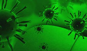
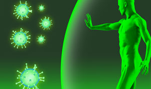
The immune system is an organization of cells, tissues[1] and organs that work together to protect (defend) our body against "foreign" invaders. Initially, these are microorganisms such as bacteria, viruses, parasites and fungi. The human body is an ideal environment for many microorganisms, which is why they try to get into it. The job of the immune system is to keep them away and, failing that, to detect and destroy them. When the immune system attacks the wrong target or is damaged (ineffective), it can trigger various diseases, including allergies, arthritis and AIDS.

The immune system is astonishingly complex. He can recognize and remember millions of different enemies, he can produce various substances and cells to mark (match) or erase each of them. The secret of its success is its developed and dynamic communication network. Billions of cells are organized into subgroups and clusters, like swarms of bees circling a hive, passing information back and forth. When the cells of the immune system receive an alarm signal, they undergo a significant change and begin to produce strong chemicals. These substances allow cells to regulate their own growth and behavior. They recruit their allies and direct them to places where the problem occurred.


[1] tissue – a group of similar cells joined together to perform the same function.

Host versus Alien

The key to a healthy immune system is its unique ability to distinguish between the body's own cells - its own and non-self - foreign cells. The body's defenses usually co-exist peacefully with cells that have their own recognition molecules. But as soon as they encounter cells or microorganisms carrying the "alien" marker, they quickly attack.

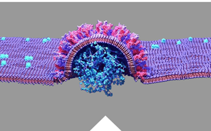
Anything that can trigger such an immune response is referred to as an antigen. The antigen may be a microorganism, such as a virus, or even a fragment of a bacterium. Tissues or cells from another person (except identical twins) therefore having the designation "foreign" acting as antigens. This explains why a transplant of "foreign" tissue may be rejected.

In certain abnormal situations, the immune system may confuse "self" and "foreign" and launch an attack against its own cells or tissues. This phenomenon (situation) is called an autoimmune disease. For example, certain forms of joint disease and diabetes are autoimmune diseases. In other situations, the immune system may react to foreign, seemingly harmless substances, such as pollen of some plants. The result is an allergy, and this type of antigen is called an allergen.

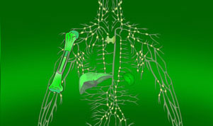
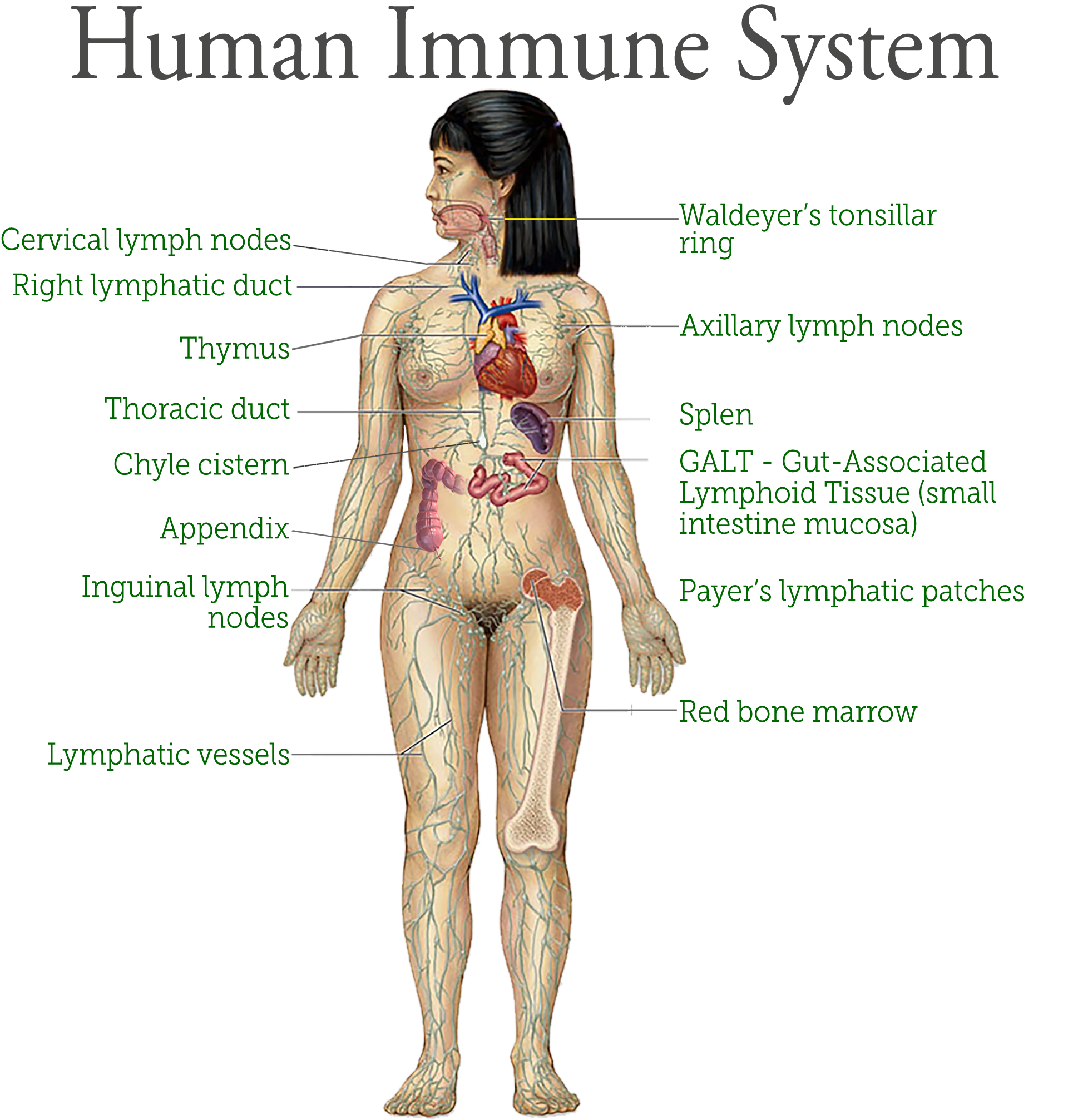
The structure of the immune system

The immune system is the body’s tool for preventing or limiting infection. Its very complex network of cells, organs, proteins, and tissues enable the immune system to defend the body from pathogens.

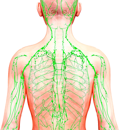
The organs (components) of the immune system are distributed throughout the body. They are called lymphoid organs because they are the home of lymphocytes – tiny white blood cells (blood cells) that are key players in the immune system.


Bone marrow

Bone marrow it is the soft tissue inside the marrow cavity of bones, is the ultimate source of all blood cells, including white blood cells destined for immune system cells. Bone marrow makes more than 220 billion new blood cells every day. Most blood cells in the body develop from cells in the bone marrow.

Bone marrow is either red or yellow, depending upon the preponderance of hematopoietic (red) or fatty (yellow) tissue. In humans the red bone marrow forms all of the blood cells with the exception of the lymphocytes, which are produced in the marrow and reach their mature form in the lymphoid organs. Red bone marrow also contributes, along with the liver and spleen, to the destruction of old red blood cells. Yellow bone marrow serves primarily as a storehouse for fats but may be converted to red marrow under certain conditions, such as severe blood loss or fever. At birth and until about the age of seven, all human marrow is red, as the need for new blood formation is high. Thereafter, fat tissue gradually replaces the red marrow, which in adults is found only in the vertebrae, hips, breastbone, ribs, and skull and at the ends of the long bones of the arm and leg; other cancellous, or spongy, bones and the central cavities of the long bones are filled with yellow marrow.

Thymus

Thymus is a pyramid-shaped lymphoid organ that, in humans, is immediately beneath the breastbone at the level of the heart. The organ is called thymus because its shape resembles that of a thyme leaf. The primary function of the thymus is to facilitate the maturation of lymphocytes known as T cells, or thymus-derived cells, which determine the specificity of immune response to antigens (foreign substances) in the body.

The thymus gland is most active during childhood. Your thymus actually starts making T-cells before you’re born. It keeps producing T-cells and you have all the T-cells you need by the time you reach puberty. After puberty, your thymus gland slowly starts to decrease in size and is replaced by fat.

Lymph nodes

Lymph nodes are located throughout the body. They are small, bean-shaped glands that play a crucial role in the immune system. During an infection, a person may notice swollen lymph nodes. The body contains hundreds of lymph nodes. They form clusters around the body and are particularly prominent in areas such as the neck, armpit and groin and behind the ears.

Lymph nodes filter lymphatic fluid, which helps rid the body of germs and remove waste products. The body’s cells and tissues dispose of waste products in lymphatic fluid, which lymph nodes then filter. During this process, they catch bacteria and viruses that could harm the rest of the body.

Lymph nodes are an essential part of the body’s immune system. Due to their function, they come into contact with toxins, which can cause them to swell. Although swollen lymph nodes are common, they may occasionally indicate lymph node cancer, or lymphoma.

Spleen

Spleen is an organ of the lymphatic system located in the left side of the abdominal cavity under the diaphragm, the muscular partition between the abdomen and the chest. In humans it is about the size of a fist and is well supplied with blood. As the lymph nodes are filters for the lymphatic circulation, the spleen is the primary filtering element for the blood. The organ also plays an important role in storing and releasing certain types of immune cells that mediate tissue inflammation.

The white pulp of the spleen contains typical lymphoid elements, such as plasma cells, lymphocytes, and lymphatic nodules, called follicles in the spleen. Germinal centres in the white pulp serve as the sites of lymphocyte production. Similar to the lymph nodes, the spleen reacts to microorganisms and other antigens that reach the bloodstream by releasing special phagocytic cells known as macrophages. Splenic macrophages reside in both red and white pulp, and they serve to remove foreign material from the blood and to initiate an immune reaction that results in the production of antibodies.

Lymphoid tissue

Lymphoid tissue consists of many organs that play a role in the production and maturation of lymphocytes in the immune response. Lymphoid tissue may be primary or secondary depending upon its stage of lymphocyte development and maturation. Secondary lymphoid tissue is collections of lymphoid tissue associated with many organs. The secondary lymphoid tissues consist of lymph nodes, tonsils, Peyer’s patches, spleen, adenoids, skin, and mucosa-associated lymphoid tissue (MALT). Mucosa-associated lymphoid tissue types are listed below:

  • GALT (gut-associated lymphoid tissue. Peyer's patches are a component of GALT found in the lining of the small intestines.)
  • BALT (bronchus-associated lymphoid tissue)
  • NALT (nasal-associated lymphoid tissue)
  • CALT (conjunctival-associated lymphoid tissue)
  • LALT (larynx-associated lymphoid tissue)
  • SALT (skin-associated lymphoid tissue)
  • VALT (vulvo-vaginal-associated lymphoid tissue)
  • TALT (testis-associated lymphoid tissue)

MALT is populated by lymphocytes such as T cells and B cells, as well as plasma cells, dendritic cells and macrophages, each of which is well situated to encounter antigens passing through the mucosal epithelium. In the case of intestinal MALT, M cells are also present, which sample antigen from the lumen and deliver it to the lymphoid tissue. MALT constitute about 50% of the lymphoid tissue in human body. Immune responses that occur at mucous membranes are studied by mucosal immunology.

Immune system cells and their products

The immune system accumulates a huge arsenal of cells, not only lymphocytes but also phagocytic cells and their relatives capable of phagocytosing (eating) cells. Certain immune cells catch all invaders, while others are trained on specific targets. To function effectively, most cells need cooperation with their companions. Sometimes immune cells communicate through direct physical contact, sometimes by releasing chemical messengers (transmitters).

The immune system collects just a little of each type of different cell needed to recognize millions of possible enemies. But when a hostile antigen appears, that "handful" of matching cells will multiply into a full army. When their work is finished, most of them will disappear and only a few will remain at the post to guard our body against future attacks.

All immune cells start as immature bone marrow stem cells. They respond to various cytokines and other signals to mature into particular ones immune cells such as T or B lymphocytes or phagocytes. Stem cells are not predetermined as to their future development and may constitute an interesting possibility in the treatment of immune system disorders. Currently, it is being investigated whether a person's own stem cells could be used to regenerate the damaged immune response in autoimmune diseases and immunodeficiencies.

B cells (B Lymphocytes)

Lymphocytes B and T are the two main types of lymphocytes. B cells work mainly by secreting substances called antibodies into body fluids. Antibodies capture antigens circulating in the blood. However, they do not have the ability to penetrate cells. The task of attacking target cells – either virus-infected cells – or cancer-infested cells is left to T cells or other immune cells (described below).

Each B cell is programmed to produce one specific antibody. For example, one B cell produces an antibody that blocks a virus that causes the common cold, while another produces an antibody that attacks a bacterium that causes pneumonia.

If a B cell encounters a trigger antigen, it causes the growth of many large cells known as plasma cells. Each plasma cell is a specialized antibody factory. Each plasma cell is a descendant of a B cell and produces millions of identical antibodies that are released into the blood. The antibody fits the antigen like a key fits a lock. Some fit very precisely, others more like a pick. But wherever an antibody and an antigen come together, the antibody marks the antigen for destruction. Antibodies belong to a family of large molecules known as immunoglobulins. Different types of antibodies perform different functions in the immune defense strategy.

IgA

Immunoglobulin A has a critical role in immune defense particularly at the mucosal surfaces. IgA is found in many body secretions, including tears, saliva, respiratory and intestinal secretions, and colostrum (the first milk produced by lactating mothers). Very little IgA is present in the serum. IgA is produced by B cells located in the mucous membranes of the body. Two molecules of IgA are joined together and associated with a special protein that enables the newly formed IgA molecule to be secreted across epithelial cells that line various ducts and organs. Although IgG is the most common class of immunoglobulin, more IgA is synthesized by the body daily than any other class of antibody. However, IgA is not as stable as IgG, and therefore it is present in lower amounts at any given time.

IgG

Immunoglobulin G is the most common class of immunoglobulin. It is present in the largest amounts in blood and tissue fluids. Each IgG molecule consists of the basic four-chain immunoglobulin structure. There are four subclasses of IgG, each with minor differences in its H chains but with distinct biological properties. IgG is the only class of immunoglobulin capable of crossing the placenta; consequently, it provides some degree of immune protection to the developing fetus. These molecules also are secreted into the mother’s milk and, once they have been ingested by an infant, can be transported into the blood, where they confer immunity.

IgM

Immunoglobulin M is the first class of immunoglobulin made by B cells as they mature, and it is the form most commonly present as the antigen receptor on the B-cell surface. When IgM is secreted from the cells, five of the basic Y-shaped units become joined together to make a large pentamer molecule with 10 antigen-binding sites. This large antibody molecule is particularly effective at attaching to antigenic determinants present on the outer coats of bacteria. When this IgM attachment occurs, it causes microorganisms to agglutinate, or clump together.

IgE

Immunoglobulin E is made by a small proportion of B cells and is present in the blood in low concentrations. Each molecule of IgE consists of one four-chain unit and so has two antigen-binding sites, like the IgG molecule; however, each of its H chains has an extra constant domain (CH4), which confers on IgE the special property of binding to the surface of basophils and mast cells. When antigens bind to these attached IgE molecules, the cell is stimulated to release chemicals, such as histamines, that are involved in allergic reactions. IgE antibodies also help protect against parasitic infections.

IgD

Immunoglobulin D molecules are present on the surface of most, but not all, B cells early in their development, but little IgD is ever released into the circulation. It is not clear what function IgD performs, though it may play a role in determining whether antigens activate the B cells.

T cells (T lymphocytes)

Unlike B cells, T cells do not recognize freely circulating antigens. Rather, their surface has specialized receptors (so-called antibody-like receptors) that recognize fragments of antigens on the surface of infected or cancer cells. T cells participate in immune defense in two main ways: some direct and regulate the immune response; others attack directly infected or cancer cells.

T helper lymphocytes (Th cells) coordinate immune responses by communicating with other cells. Some stimulate nearby B cells to produce antibodies, others call on phagocytes, and others activate other T cells.

Tc cells

Cytotoxic T lymphocytes, also called Tc cells or Tc lymphocytes (Cytotoxic T Lymphocytes - CTLs), perform various tasks. These cells directly attack other cells that carry certain foreign or abnormal molecules on their surface. Cytotoxic T cells are particularly useful in defense against viruses because viruses are often hidden from other elements of the immune system because viruses often multiply inside infected cells. These cells can detect even small fragments of the virus protruding through the cell membrane of a virus-infected cell and attack it to destroy it.

In most cases, T cells can only recognize an antigen if it is carried on the cell surface by its own MHC molecules (major histocompatibility complex). MCH molecules are proteins recognized by T cells when they distinguish between self and foreign. Own MHC molecules create a recognizable scaffold for presenting foreign antigens to T cells.

Although MHC molecules are needed by Tc cells to attack foreign invaders, they pose problems in the case of organ transplantation. Each cell of the body has MHC proteins on its surface, and each person has his or her own set of these proteins. If a Tc lymphocyte recognizes "foreign" MHC molecules on the surface of a cell, which happens in transplants, it will begin to destroy it. Therefore, it is necessary to select organ donors with the set of MH molecules (major histocompatibility complex) that is most similar to the recipient's set. Otherwise, Tc lymphocytes will probably attack the transplanted organ and lead to its rejection.

NK cells

NK cells, or natural killer cells, are another type of white blood cells of the lymphocyte class. Like cytotoxic T cells (LTc), NK cells are armed with granules filled with powerful chemicals. However, while killer cytotoxic T cells look for antigen fragments attached to molecules of "their" MHC, NK cells recognize cells that do not have molecules of "their" MHC. Therefore, NK cells have the ability to attack various types of foreign cells.

Both types of killer cells kill through contact. Assassins attach themselves to their target, turn their weapons on him and trigger a deadly chemical explosion.

Phagocytes and their relatives

Phagocytes are large white cells that can devour and digest microbes and other foreign particles. Monocytes are phagocytes that circulate in the blood. When monocytes enter the tissues, they transform into macrophages.

Specialized types of macrophages are found in many organs: lungs, kidneys, brain and liver. Macrophages perform many functions. Like cleaners, they free the body of used cells and other debris (garbage). They display pieces of foreign antigens so as to attract the attention of the appropriate lymphocytes. They project out an incredible multitude of powerful chemical signals, known as monokines, that are important for the immune response.

Another type of immune system cells are granulocytes. They contain granules filled with powerful chemicals that allow granulocytes to destroy microorganisms. Some of these substances, such as histamine, are also involved in the processes of inflammation and allergy.

One of the granulocytes, the neutrophil, is also a phagocyte; it uses previously stored chemicals to break down and digest microbes. Eosinophils and basophils are other granulocytes that release chemicals from their granules, spraying them onto nearby microbes and harmful cells.

Mast cells are the twins of basophils, except that they are not blood cells. They are found in the tissues: lungs, skin, tongue, mucous membrane lining the nasal cavity and the digestive tract, where they are responsible for allergy symptoms.

A related structure is platelets. These are cell fragments that also contain granules. The function of platelets related to blood cell aggregation, coagulation and wound healing also includes immune activation.

Cytokines

Cytokines are proteins that function as chemical messengers in your immune system. Your immune system is a network with several parts that work together to protect your body from threats, like germs that can make you sick. It contains immune cells that fight invading pathogens (like viruses and bacteria), allergens and other harmful substances that enter your body. Cytokines signal those immune cells to fight the invaders. Even when there’s no threat, cytokines send signals to other cells that keep your immune system functioning.

The components of the immune system communicate with each other by exchanging chemical messengers called cytokines. These proteins are secreted by cells and act on other cells to coordinate an appropriate immune response. Cytokines include a diverse range of interleukins, interferons, and growth factors. Some cytokines are types of chemical switches that turn certain types of immune cells on or off.

Different types of cytokines

Cytokines include different types of proteins that tell immune cells where to go and what to do to keep your immune system functioning correctly.

  • Chemokines direct immune cells toward places in your body where they can fight infection.
  • Interferons signal cells to put up their defenses against viruses invading your body. In this way, interferons “interfere” in the process that allows viruses to replicate, or make more viruses once they’ve invaded a healthy cell.
  • Interleukins get their name from “inter” which means between and “leukocyte,” which is another name for a white blood cell. Originally, scientists thought that leukocytes alone released interleukins and only relayed messages to other leukocytes. But now we know that cells other than leukocytes release these proteins. Also, interleukins can relay messages between cells that aren’t leukocytes.
  • Tumor necrosis factor (TNF) helps regulate inflammation in your body. TNF also signals to immune cells that kill tumor cells.
  • Colony-stimulating factors CSF signals hematopoietic stem cells to develop into specific cell types. Hematopoietic stem cells (HSC) are precursor cells that give rise to all blood cell types: white blood cells, red blood cells and platelets. These changes take place during a process called hematopoiesis. For example, granulocyte-colony stimulating factor (G-CSF) signals an HSC to become a white blood cell called a neutrophil. Neutrophils help fight infection.
Some cytokines get their names from the type of cell that makes them, including:
  • Lymphokines – produced by lymphocytes, a type of white blood cell.
  • Monokines – produced by monocytes, a type of white blood cell.

One of the cytokines, interleukin 2 (IL-2), triggers the production of T cells by the immune system. The immune-stimulating properties of IL-2 have traditionally made it hopeful in the treatment of a number of diseases. Clinical trials are underway testing their benefits in other diseases such as cancer, hepatitis C, HIV infection and AIDS. Other cytokines are also being investigated for their potential therapeutic benefits and clinical applications.

Other cytokines chemically attract particular types of cells. These so-called chemokines are released by cells at the site of damage (injury) or infection and call other immune cells to the site to help repair the damage and fight off invaders. Chemokines often play a key role in inflammation and are promising targets for new drugs that regulate the immune response.

The complemment system

The complement system contains approximately 25 proteins that work together to "complement" the action of antibodies in destroying bacteria. Complement also supports the body's release from antigen-antibody complexes. Complement proteins, which cause blood vessels to dilate and cause their permeability, are involved in the development of redness, swelling, pain, increase in temperature and loss of function that characterize the inflammatory response.

Complement proteins circulate in the blood in an inactive form. When the first complement protein is activated – typically by an antibody that locks with an antigen – a domino effect is triggered. Each component (takes) contributes to a precise chain of steps known as the complement cascade. The end product is a cylinder that sticks into the cell wall, creating a hole (hole) in it. Fluids and particles flow in and out through it into the cell, which swells (buzzes) and bursts. Other components of the complement system make bacteria susceptible to phagocytosis and/or attract other cells to the region.

Structure of the Immune System

The immune system is a very complex structure consisting of cells, tissues and organs that work together to protect (defend) our body against "foreign" invaders . . .

How the Immune System Works?

It is one of nature's most fascinating inventions. It easily protects us against billions of bacteria, viruses and pathogens. We don't realize that the immune system . . .

Immunodeficiencies

Immunodeficiencies are disorders of the immune system that are characterized by a reduced or lack of ability to . . .

Maintain Healthy Immune System

If we are not dealing with an immune system disease, it is usually enough to use a few simple . . .

News

The European Medicines Agency (EMA) has approved gene therapy for the treatment of severe combined immunodeficiency due to adesine deaminase deficiency (ADA-SCID), which is the result of a genetic mutation - reports New Scientist. You can read about gene therapy, what it is and its prospects, on the website News Medical Life Sciencies

17.01.2024

Source: Memorial Sloan Kettering Cancer Center Library / LibGuides / COVID Impacts / Immune Dysfunction

Detailed information and resources on the long-term health consequences of COVID-19 infection and the broad social impacts of the COVID-19 pandemic.

One of the most concerning long-term effects of COVID-19 is the dysregulation and dysfunction of the immune system.

08.08.2023

Source: Nutrients Authors: van Steenwijk H, Bast, A and de Boer A.

Beta-glucans derived from mushrooms are bioactive long-chain polysaccharide compounds, insoluble in water and with immunomodulatory properties. Knowledge of the action and functions of beta-glucans, which have been used in traditional medicine for centuries, is developing thanks to modern immunological and biotechnological methods.

04.08.2023

Source: Oncology Reports; Authors: Hiromi Okuyama Akira Tominaga, z Laboratory of Immunology, Faculty of Pharmacy, Osaka Ohtani University, Tondabayashi, Osaka 584-8540, Japan

Spirulina lipopolysaccharides inhibit tumor growth in a Toll-like receptor 4-dependent manner by altering the cytokine milieu from interleukin-17/interleukin-23 to interferon-γ